How Does The Ventricle Generate Pressure And Flow?
This section will provide a way to understand how events that occur on the cellular level (excitation-contraction) translate into a functional fluid pump.
|
Relationships Between Pressure, Flow, Volume,
and Compliance
|
1. Blood flows because of pressure gradients
|
2. Pressure is determined by the Volume in a chamber and the
Compliance of the chamber wall
|
3. Volume is determined by the net result of blood entering versus
leaving the chamber
|
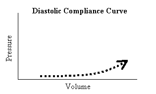
First, let's look at the passive propeties of the ventricle. The ventricle is in this passive state during diastole, prior to excitation-contraction.
| 1. The pressure in the ventricle is less than the venous pressure, so blood flows into the ventricle. |
 |
| 2. The ventricular wall is very compliant, so the volume entering does not cause much of an increase in pressure. (Some diseases cause the ventricle to be less compliant. |
 |
| 3. As the wall gets further stretched, it becomes less compliant, so there is a more pronounced rise in pressure for a given change in volume. |
 |
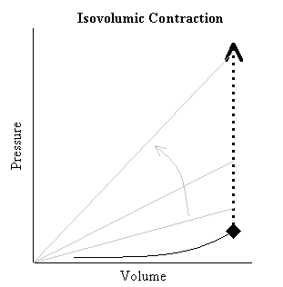
| 4. Next, two events occur: (1) a wave of excitation-contraction occurs on the cellular level throughout the myocardium, and (2) the pressure in the ventricle rises with NO change in volume (i.e. Isovolumic phase). So how does #1 cause #2? Answer: The actin-myosin cross-bridging result in a decrease in the compliance of the myocardium. Note: while the term COMPLIANCE refers to volume divided by pressure, the term ELASTANCE, E is the inverse relationship, pressure divided by volume (P/V). So, during isolvolumic contraction, Ventricular Elastance is increasing. This should be obvious, since P is increasing, while V remains constant.
This also can be appreciated by looking at the slope of the lines connecting the origin with any point during the isovolumic contraction phase on the P-V loop (see the grey lines).
|
 |
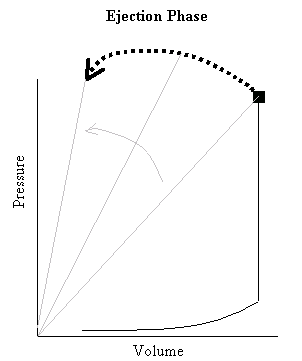
| 5. Next, comes the ejection phase. During this phase, ventricular volume is decreasing (volume is being ejected out across the aortic valve into the system vasculature), while pressure is rising a bit, and then falling. Since both terms (P and V) are changing, it may not be as clear what is happening to Ventricular Elastance, but by looking at the P-V loop, it should be clear the Elastance continues to increase during the ejection phase. From the beginning of systole (the end-diastolic diamond in the above Figure), until the last moment of systole (the arrow head in the Figure on the right), ventricular elastance continues to increase. The changes that occur on the cellular level serve to change the mechanical properties of the muscle, namely, the muscle becomes more stiff. |
 |
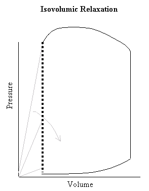
| 6. Lastly, when systole terminates, there is active de-coupling of actin and myosin cross-bridges. This is associated with the isolvolumic relaxation phase. Since pressure is rapidly declining while ventricular volume remains constant, it should be obvious that the reason the pressure is declining is due to a decrease in Ventricular Elastance. In order to get a feel for the dynamic nature of cardiac contraction and the pressure volume loop, play this quick time movie below which shows the loop in "real time.". The slope of the white line is Elastance.
|
 |
|

