Building a Better Gross Anatomy Dissector with iBooks
Three years ago, medical students from Columbia University’s College of Physicians & Surgeons approached Gross Anatomy Course Director Professor Paulette Bernd, Ph.D. with an ambitious idea: to create a new dissector for the course. The group ended up creating an innovative text from scratch, taking their own photographs and writing clearer, simpler instructions for dissecting a cadaver.
Not long after, Dr. Bernd mentioned to Michelle Hall of the Columbia Center for New Media Teaching and Learning (CCNMTL) that she was interested in adapting the dissector for the iPad using iBooks Author. Hall was enthusiastic and arranged for CCNMTL to adapt one of her chapters to create a prototype. Dr. Bernd took it from there, creating a complete ebook version. The innovative dissector has been featured in the Wall Street Journal and several on-campus publications. This spring, the dissector made its debut as five different books, each dedicated to a different region of the body, in the Apple iBooks Store.
CCNMTL recently spoke with Dr. Bernd about the process of creating an e-dissector and how it has affected teaching and learning in the Gross Anatomy course.
CCNMTL: What inspired you to make your own dissection manual?
Dr. Bernd: Gross Anatomy is one of the first courses medical students take. It’s the foundation from which everything else comes from.
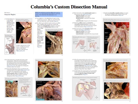
The genesis of this project began because students and faculty were dissatisfied with commercially available dissectors. Furthermore, Columbia’s new curriculum required that courses be streamlined, so an easy-to-follow dissector became a necessity given the time constraints. The old dissector was 90% text and had complicated instructions and more detailed dissections than were needed for our class. It also only had line drawings in it. The problem is that it shows you drawings of an ideal of a finalized dissection. We wanted to show step by step pictures of a real dissection.
At this time, [a photographic dissector] didn’t exist at all. I’m not one to reinvent the wheel; if there had been something out there, I would have used it. One publisher offered a dissection manual that was editable in the sense that I could edit the text, but I couldn’t add my own images. So we created it from scratch.
What was the process of creating the dissector?
This project would never have been possible without the willingness and dedication of the initial group of medical students who worked on the project: Dustin Tetzl, Justin Neira, and Jose Ramirez from the P&S Class of 2014 and Lily Grossman from the SUNY Downstate Medical School Class of 2014.
They did a dissection and took photos all along. Then they wrote up a rough draft of the text using the current book as a guideline. Then I edited and reorganized it in a way I thought made more sense for dissecting. I gave what I thought were better, simpler instructions.
That first year, the dissector was written with Microsoft Word using their publisher mode, and we printed out a hard copy that students used in the lab. A PDF was posted on CourseWorks so students could access it for study. Apple released iBooks Author right after that course ended, and I said, “This is a platform we could use.” CCNMTL helped me get started, and then I did the conversion to iBooks myself.
Students continued to work with me over the next two summers (2012 and 2013) to add photos, a glossary, quizzes, videos, etc. These students were Michael Bruno, CDM Class of 2016; Stephen Cassidy, P&S Class of 2017; Michelle Castro De Fazio, CDM Class of 2015; Adam De Fazio, P&S Class of 2015; Bryce Farr, CDM Class of 2016; Tristan Hunt, P&S Class of 2015; Colin Klenk, P&S Class of 2016; Jesse Koskey, P&S Class of 2015; Alison Levy, P&S Class of 2015; Nina Ragaz, P&S Class of 2016; and John Ryan, P&S Class of 2016.
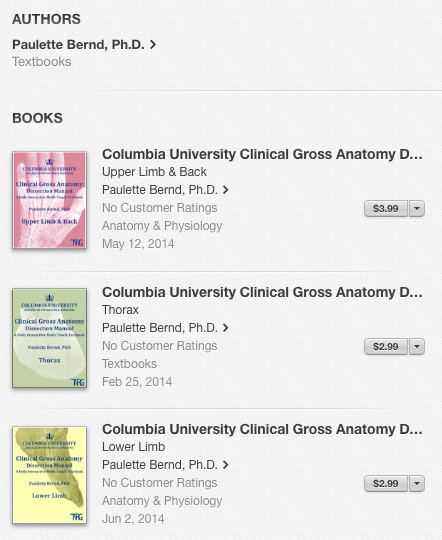 Three of the dissector books in the iBooks Store.
How do students use the iBooks dissector in the classroom?
Each table in the lab has a full-size iPad thanks to a generous donation from Diana and P. Roy Vagelos, M.D. The iPads are stored in a student-constructed case for safe keeping and storage. Students can also put the dissector on their own iPad, or, now, on a Mac laptop that runs OS Mavericks. I’m also hoping iBooks will be able to open on an Android tablet eventually.
One thing is for sure: students use this dissector. The old book, they had it open, but I found they usually were not using it or paying attention to it.
The new dissector has been really well received. Students get a lot out of it. They are much less frustrated, and they do a better job on the dissections. In addition, exam grades went up substantially the first year and have gone up a little every year thereafter.
In addition to the new dissector, how else have you changed the Gross Anatomy curriculum at Physicians & Surgeons?
My approach when developing the new Gross Anatomy curriculum was to make the course more efficient yet keep its clinical relevance. One way to do that is to emphasize imaging, so the students come out of this course with a lot of knowledge of anatomy on CT scans, MRIs, etc., because that is how most of them will be seeing anatomy, not in surgery. The students dissect one day a week, and another day a week they do a non-dissecting lab in which we look at imaging and clinical cases.
When I came here, one thing I was trying to do is reduce stress for students. All the basic science courses here are pass/fail. These are superb students. I didn’t feel it was necessary to weed out weak students. Teamwork is one of the emphases of the new curriculum. So I decided to let them take their practical exam in the lab in their dissecting group of four students rather than alone. I started doing that before I had the new dissector.
It’s really terrific, because when they take the exam together, they have to be very objective in their decision making, and they have to convince one another they are correct. We overhear some fascinating conversations.
Anatomy is one of the few courses you don’t have to convince people they have to learn. Anecdotally, students say they’re working even harder because they don’t want to let their teammates down. These groups get so bonded, they go out to dinner together years later.
What future plans do you have for the dissector and the Gross Anatomy course?
Two years ago, with the cooperation of the Radiology Department, we began to CT the cadavers. We then put each CT scan on that table’s iPad so students can refer to their “patient’s” radiologic findings as they encounter pathology in their dissection. My next goal is to make all the imaging information from the non-dissecting sessions digital.
Columbia is building a new lab gross anatomy lab in the new Medical and Graduate Education Building. In the new space, there will be a monitor at every station so students will be using the dissector on a larger screen.